


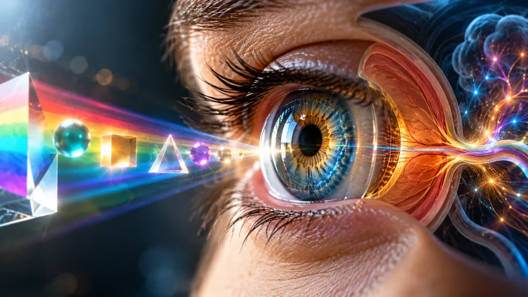

The human eye is one of the most remarkable organs in the body, allowing us to recognize colors, identify shapes, and understand the world around us. From noticing the bright colors of a sunset to recognizing familiar faces, our visual system processes an enormous amount of information every day. However, seeing involves much more than simply opening our eyes.
The human eye works by collecting light and converting it into electrical signals that the brain interprets as images. Specialized cells inside the retina detect different aspects of light, including brightness and color. The brain then processes these signals to identify objects, understand shapes, and recognize visual patterns.
Understanding how the human eye sees colors, shapes, and light requires exploring several interconnected structures. The cornea, pupil, lens, retina, optic nerve, and visual cortex all contribute to sight. This guide explains how these parts work together, how color vision develops, and how the brain transforms light into the images we experience.
How Does the Human Eye Work?
The human eye functions as a biological optical system that receives light and focuses it onto a light-sensitive layer called the retina. Light enters through the transparent cornea and passes through the pupil before reaching the lens. These structures work together to direct incoming light toward the back of the eye. The retina then begins converting light information into neural signals.
The lens changes its shape to help focus light from objects at different distances. This process is called accommodation and is controlled by muscles inside the eye. When light reaches the retina, specialized cells respond to its intensity and wavelength. The resulting signals travel through connected nerve cells toward the optic nerve.
The brain receives these signals and processes them into meaningful visual information. It identifies colors, shapes, movement, distance, and relationships between objects. Although we often think of vision as something the eyes do alone, the brain performs much of the interpretation. Clear sight depends on effective cooperation between the eyes and the nervous system.
The Main Parts of the Human Eye
The human eye contains several structures that contribute to the process of vision. The cornea is the transparent outer surface responsible for much of the eye’s focusing power. Behind it, the iris controls the size of the pupil, regulating how much light enters. The lens provides additional focusing adjustments.
The retina is a thin layer of nerve tissue located at the back of the eye. It contains specialized light-sensitive cells called rods and cones. These cells detect incoming light and begin the process of converting it into electrical activity. The optic nerve carries processed visual information from the retina toward the brain.
Other structures support normal eye function and visual clarity. The sclera forms the tough outer protective layer, while internal fluids help maintain the eye’s shape. The macula is a specialized retinal region important for detailed central vision. Together, these structures create an efficient system for collecting and processing visual information.
How Does Light Enter the Human Eye?
Vision begins when light reflected or emitted by objects travels toward the eyes. The first major structure it encounters is the cornea, a transparent curved surface that bends incoming light. This bending process is called refraction. It helps direct light toward the retina so that the visual system can form a focused image.
After passing through the cornea, light travels through the pupil, which is the opening in the center of the iris. The iris adjusts pupil size according to lighting conditions. In bright environments, the pupil usually becomes smaller to limit incoming light. In darker environments, it becomes larger to allow more light to enter.
The light then passes through the lens, which makes additional focusing adjustments. A properly focused optical image forms on the retina at the back of the eye. Retinal cells respond to the light and initiate electrical signals. These signals begin the neurological process that eventually produces conscious visual perception.
How Does the Retina Help Us See?
The retina is the light-sensitive tissue responsible for converting incoming light into neural information. It lines the inner surface at the back of the eye and contains several interconnected layers of nerve cells. Its most important light-detecting cells are rods and cones. These photoreceptors respond to light through specialized chemical reactions.
When light reaches photoreceptors, it changes the activity of light-sensitive molecules called visual pigments. This produces changes in the cells’ electrical signals through a process known as phototransduction. The information is then processed by other retinal neurons. These cells help organize visual signals before they leave the eye.
The retina does more than simply detect brightness. Its neural circuits contribute to processing contrast, spatial information, and changes in light. This early processing helps prepare visual information for interpretation in the brain. The retina therefore plays an essential role in recognizing objects, identifying colors, and detecting movement.
Rods and Cones: The Cells Responsible for Vision
Rods and cones are two types of photoreceptor cells found in the human retina. Both detect light, but they perform different functions. Rods are highly sensitive to low light levels, while cones support color vision and fine visual detail. Their combined activity allows humans to see across a wide range of lighting conditions.
Rods are especially important for vision in dim environments, such as during nighttime or in poorly illuminated rooms. They are highly sensitive to light but do not provide normal color discrimination. This explains why colors often appear faded in darkness. Rods are more numerous in peripheral retinal regions than in the central fovea.
Cones function best under brighter lighting conditions and are concentrated in the central retina. They enable detailed vision and help distinguish different colors. Humans typically have three major classes of cones with different wavelength sensitivities. Together, these cells make it possible to recognize colorful objects and observe fine visual details.
How Does the Human Eye See Colors?
The human eye sees colors by detecting different wavelengths of visible light through specialized cone cells in the retina. Visible light contains a range of wavelengths that can produce different color sensations. When light enters the eye, cones respond according to their spectral sensitivities. The brain interprets their combined activity to create color perception.
Most people have three types of cone photoreceptors, commonly called S, M, and L cones. These are most sensitive to shorter, medium, and longer wavelengths of visible light, respectively. Each cone type responds to a range of wavelengths rather than detecting only one color. Color perception depends on comparing the signals produced by these different cells.
For example, light from a red object usually produces a pattern of cone responses different from the pattern produced by a blue object. The visual system compares these signals and interprets them as different colors. Lighting conditions, surrounding colors, and brain processing also influence perception. Color vision is therefore a combined function of the eyes and brain.
Why Do We See Different Colors?
Objects appear to have different colors because they interact with visible light in different ways. When light reaches an object, some wavelengths may be absorbed while others are reflected or transmitted. The light that eventually enters our eyes influences the color we perceive. This process explains why materials can appear red, blue, green, or other colors.
A green leaf, for example, generally reflects more light from certain green portions of the visible spectrum than from some other wavelengths. The reflected light stimulates cone cells in the retina. The brain processes the resulting signals and produces the sensation of green. The exact appearance also depends on illumination and surrounding visual conditions.
Color perception can change when the same object is viewed under different lighting conditions. A surface may appear slightly different in daylight compared with artificial indoor lighting. The brain partially compensates for these changes through a process called color constancy. This ability helps us recognize familiar object colors across varying environments.
How Does the Human Eye See Shapes?
The human eye recognizes shapes by detecting differences in light intensity, contrast, edges, and spatial patterns. When light from an object reaches the retina, it creates a distribution of activity across photoreceptor cells. Retinal neurons begin processing this information. The brain then identifies boundaries and organizes visual signals into meaningful forms.
Edges are particularly important for recognizing shapes because they indicate changes between neighboring regions. For example, the boundary between a dark object and a bright background produces a contrast pattern. Specialized neurons in the visual system respond to these differences. Their activity helps distinguish an object’s outline from its surroundings.
The brain combines information about edges, angles, curves, and spatial relationships to recognize familiar objects. This process allows people to identify circles, squares, faces, and complicated three-dimensional structures. Previous visual experience also contributes to recognition. Shape perception is therefore the result of both incoming sensory information and neural interpretation.
How Does the Brain Process Visual Information?
The brain plays a central role in turning electrical signals from the retina into conscious visual experiences. Information travels through the optic nerves and important processing regions before reaching the visual cortex. The primary visual cortex is located in the occipital lobe near the back of the brain. It begins processing important features of the incoming signals.
Different visual pathways contribute to recognizing objects, movement, color, and spatial relationships. Some processing regions help identify what an object is, while others contribute to understanding its location and movement. These pathways communicate extensively rather than operating as completely separate systems. Their coordinated activity creates a unified visual experience.
The brain also uses attention, memory, and previous experience when interpreting what we see. Familiar objects can often be recognized quickly because the visual system has learned their patterns. This explains why perception involves more than recording light. The brain continuously organizes and interprets sensory information to help us understand our surroundings.
How Do We See Objects at Different Distances?
The eye focuses on objects at different distances through a process called accommodation. This process involves changes in the shape of the lens controlled by the ciliary muscles. When viewing nearby objects, the lens becomes more rounded to increase focusing power. When viewing distant objects, the lens becomes relatively flatter.
The cornea provides most of the eye’s basic focusing power, while the lens makes additional adjustments. Together, they direct light toward the retina to create a clear optical image. If the light does not focus correctly, the image may appear blurry. This can occur because of differences in eye shape or focusing ability.
Common refractive errors include nearsightedness, farsightedness, and astigmatism. These conditions affect how light is focused on the retina. Eyeglasses, contact lenses, and certain medical treatments can help correct focusing problems. The appropriate option depends on the individual’s visual needs and professional eye examination findings.
How Does the Human Eye See in Darkness?
Seeing in darkness depends largely on rod photoreceptors, which are especially sensitive to low levels of light. In dim environments, rods become more important because cone cells require stronger illumination for their best performance. This allows people to detect objects and movement even when lighting is limited.
When moving from a bright environment into darkness, vision does not immediately reach its maximum sensitivity. The visual system gradually adapts through changes in photoreceptor activity and visual pigment availability. This process is called dark adaptation. Full adaptation can take several minutes or longer, depending on the circumstances.
Colors usually appear less vivid in low light because cone activity becomes less effective. Peripheral vision may also become especially useful for detecting faint objects. However, human night vision has limitations, and extremely low light can prevent clear visual recognition. Adequate illumination remains important for activities that require precision and safety.
How Do Both Eyes Work Together?
Humans normally use two eyes that view the world from slightly different positions. Each eye receives its own optical image and transmits visual information to the brain. These images contain small differences because the eyes observe objects from separate angles. The brain combines information from both eyes to support a unified visual experience.
Binocular vision helps improve depth perception and the ability to judge distances. The brain analyzes differences between the two retinal images, a process known as binocular disparity. This information contributes to stereoscopic depth perception. Additional visual cues, such as motion and perspective, also help estimate distance.
Eye coordination is important for keeping both eyes directed toward the same object. Muscles surrounding the eyes control their movements and alignment. When the eyes work together effectively, the visual system can interpret spatial information more accurately. Problems with alignment or coordination may affect comfort, depth perception, or image clarity.
Why Do We See an Image Upside Down on the Retina?
The cornea and lens form a real optical image on the retina that is inverted relative to the outside world. Light rays from the upper portion of an object are focused onto lower retinal regions. Rays from the lower portion reach upper retinal regions. This arrangement results from the optical properties of the eye’s focusing structures.
Despite the inverted retinal image, people experience the world in its familiar upright orientation. The brain does not simply rotate a photograph inside the head. Instead, it interprets visual signals using learned relationships between sensory information, body position, and the environment. Perception develops through coordinated neural processing.
This demonstrates why vision cannot be explained by the eye alone. The retina provides structured information, while the brain interprets spatial relationships and object positions. Our visual experiences depend on how the nervous system processes these signals. The apparent orientation of the world is part of this broader perceptual system.
What Causes Color Blindness?
Color vision deficiency, often called color blindness, occurs when the visual system has difficulty distinguishing certain colors. The most common forms involve differences in the function of cone photoreceptors. Red-green color vision deficiencies are particularly common. Complete inability to perceive color is much less common.
Many color vision deficiencies are inherited and associated with genetic differences affecting cone pigments. Other forms can develop because of eye diseases, injuries, medications, or changes involving the nervous system. The specific colors that are difficult to distinguish depend on the type of deficiency. Experiences vary considerably between individuals.
Eye care professionals can evaluate color vision using specialized tests designed to identify common deficiencies. Recognizing the condition can help individuals understand certain visual challenges. Many people with color vision differences manage everyday activities successfully. However, some occupations or tasks may require particular color discrimination abilities.
How Does the Eye Adjust to Bright Light?
The human eye adjusts to bright environments through several protective and adaptive mechanisms. One visible response is pupil constriction, in which the iris makes the pupil smaller. This reduces the amount of light entering the eye. The response helps regulate retinal illumination when surrounding brightness increases.
Photoreceptor cells also adjust their sensitivity according to light levels. In bright conditions, cone cells support detailed vision and color discrimination. The visual system adapts to changes in illumination so that objects remain recognizable across different environments. These adjustments help maintain useful vision despite substantial variations in brightness.
However, the eye’s natural responses do not provide complete protection against intense light sources. Looking directly at the Sun can damage the retina, even when discomfort is limited. Appropriate eye protection is important in hazardous environments. Normal adaptation mechanisms should not be considered substitutes for recommended protective measures.
What Factors Affect Human Vision?
Human vision depends on several biological and environmental factors, including eye health, lighting conditions, age, and neurological function. Problems involving the cornea, lens, retina, or optic nerve can affect visual clarity. Changes in any of these structures may influence how colors, shapes, and movement are perceived.
Age-related changes can also affect focusing ability and sensitivity to light. For example, the lens gradually becomes less flexible with age, making nearby objects more difficult to focus on. Other conditions may affect contrast sensitivity or color perception. Regular eye examinations can help identify visual changes and underlying health concerns.
Environmental factors such as glare, poor lighting, and prolonged screen use can influence visual comfort. Dryness or temporary focusing fatigue may make vision feel uncomfortable during certain activities. Appropriate lighting and regular breaks can help improve comfort. Persistent changes in vision should be evaluated by a qualified eye care professional.
How to Keep Your Eyes Healthy
Maintaining eye health involves a combination of healthy habits, appropriate protection, and regular professional care. Balanced nutrition supports overall health, including the normal function of eye tissues. Adequate sleep and good management of relevant medical conditions can also contribute to visual well-being. These habits help support healthy eye function throughout life.
Protecting the eyes from harmful ultraviolet radiation is another important consideration. Sunglasses that provide appropriate UV protection can reduce exposure during outdoor activities. Protective eyewear is also valuable when participating in activities that could cause eye injuries. Following relevant safety recommendations helps reduce preventable harm.
Regular eye examinations can identify vision problems and certain eye conditions before noticeable symptoms develop. People experiencing sudden visual changes, eye pain, or unusual symptoms should seek appropriate medical attention. Healthy habits cannot prevent every eye condition, but they support overall visual health. Professional assessment remains important when vision problems occur.
Conclusion
The human eye sees colors, shapes, and light through a coordinated process involving optical structures, specialized retinal cells, and the brain. Light enters through the cornea and pupil before being focused by the lens onto the retina. Rods and cones convert light into neural signals. The brain then interprets this information as meaningful visual experiences.
Color vision depends on different cone photoreceptors responding to wavelengths of visible light, while shape recognition involves contrast, edges, and spatial patterns. Rods support vision in dim environments, and the lens adjusts focus for objects at different distances. Both eyes also work together to support depth perception and spatial awareness.
Understanding how the human eye works reveals the complexity of everyday vision. Clear sight depends on healthy eye structures, effective neural communication, and the brain’s ability to process visual information. Protecting eye health through appropriate care and regular examinations helps maintain this remarkable sensory system.
Frequently Asked Questions
How Does the Human Eye See Colors?
The eye sees colors through three types of cone cells in the retina. These cells respond differently to visible light wavelengths, and the brain compares their signals to produce color perception.
How Does the Human Eye Recognize Shapes?
The visual system identifies shapes by processing edges, contrast, curves, and spatial patterns. The retina detects light differences, while the brain combines this information to recognize objects and their outlines.
What Is the Role of the Retina in Vision?
The retina contains light-sensitive rod and cone cells that convert incoming light into neural signals. These signals pass through retinal circuits and travel toward the brain for further processing.
Why Can We See Better in Bright Light Than in Darkness?
Bright light allows cone cells to function effectively, supporting sharp detail and color vision. In darkness, rods become more important, but color perception and visual detail are generally reduced.
Which Part of the Brain Controls Vision?
The occipital lobe contains the primary visual cortex, which processes important visual information. Other connected brain regions help interpret colors, shapes, movement, depth, and object recognition.